Introduction
Tuberculosis (TB) is a highly contagious disease caused by Mycobacterium tuberculosis, which currently threatens a significant proportion of the world population, mainly due to its ability to induce latent infection. It has been estimated that there are approximately nine million of new cases and 1.4 million deaths due to the disease each year, ranking tuberculosis second in mortality of all infectious diseases worldwide 1. Upon infection, active TB develops in 5% of individuals while 90% carry a latent infection for the rest of their lifetimes, as they are unable to fully eradicate the pathogen 2,3. During the encounter with the host, the bacilli enter the lungs in aerosolized particles infecting and activating alveolar macrophages and dendritic cells. Infected macrophages release cytokines and chemokines triggering a strong immune inflammatory response that leads to the formation of a granuloma, an organized cellular accumulation around the bacilli 4. It has been suggested that the host immune response is able to adjust and respond to the physiological state of the bacterium modulating the expression of genes directly in the site of infection 5. Complete eradication of the bacilli does not take place, since the bacterium has developed strategies not only to persist within the granuloma for long term survival, but also to exploit it for local and systemic dissemination 6. Thus, under certain physiological (malnutrition, aging, etc.) or pathological (HIV infection, diabetes, cancer, etc.) conditions, M. tuberculosis is able to reactivate and escape from the granuloma and disseminate 7. A better understanding of the mechanisms involved in granuloma formation and maintenance may help in the development of targeted therapies against tuberculosis 4.
Different animal 8,9 and human in vitro models 10-12, have been developed to unravel the complex sequence of early molecular events involved in granuloma formation. In vitro models in particular represent a valuable tool for the identification of the molecular mechanisms implicated in the early immune response to defined mycobacterial cells 13. In the present study we used the in vitro model for granuloma development proposed and characterized by Puissegur et al. 11, which proved to be useful to study the molecular interactions between mycobacteria and human host cells using live mycobacteria and peripheral blood mononuclear cells (PBMCs). Using this model we determined global gene expression profiles induced during in vitro formation of granulomas in response to M. tuberculosis H37Ra strain. Analysis of genes and pathways altered during development of these in vitro granulomas holds the potential to aid in the understanding of early molecular events involved in this host-microbe interaction.
Materials and Methods
Immune cells and bacteria
Peripheral blood was obtained from healthy donors after they signed informed consent documents. Peripheral blood mononuclear cells (PBMCs) were isolated using gradient centrifugation on Ficoll-Hipaque 1077 (Sigma Chemical Co., St Louis MO, USA) and suspended in RPMI 1640 supplemented with 10% fetal bovine serum. M. tuberculosis (H37Ra) cells were cultured on modified Lowenstein-Jensen Medium Base. Bacteria were collected in Middlebrook 7H9 Broth (BD Difco Biosciences, Mountain View, CA, USA) and thoroughly mixed with syringe needles. The bacteria were cultured with a serial dilution on modified Lowenstein-Jensen medium and the viability was monitored by counting the colony-forming units (CFU).
Induction of in vitro granulomas
Peripheral blood mononuclear cells were transferred into 24 well tissue culture plates at a concentration of 1×10⁵ cells per well in RPMI 1640 with 10% FBS. Freshly prepared M. tuberculosis H37Ra or BCG cells were subsequently added to each well with a multiplicity of infection (MOI) of 0.1 based on trial results with different MOIs. The cells were cultured for periods from 24 h to 5 days at 37° C with medium changed every other day. To assess the specificity of the granuloma reaction, PBMCs were also cultured in the presence of Escherichia coli ATCC 25922 or Staphylococcus aureus ATCC 25923, with a MOI of 0.1. Peripheral blood mononuclear cells cultured in the absence of bacteria were also included as controls.
Light microscopy and cell examination
To monitor the progress of cellular aggregation, cultured cells were observed under an inverted microscope (Nikon, Chiyoda-ku, Tokyo, Japan) and photographs were taken with a Nikon capture system. Cells were stained with Wright-Giemsa (W-G) modified staining (Sigma-Aldrich, St Louis, MO, USA) according to the manufacturer's instructions every 24 h up to 5 days of culture.
Transmission electron microscopy
At 48 h post-infection, cellular aggregations were carefully collected, fixed for 4 h at 4o C in 2% glutaraldehyde in 0.1 M cacodylate buffer with 6 mM CaCl2, pH 7.4. After washing with cacodylate buffer, fixed granulomas were treated for 1 h with 1% osmium tetroxide in 0.1 M cacodylate buffer, dehydrated and embedded in an Epon-araldite resin. Sections of 0.5 µm were obtained on a microtome and mounted on copper grids, stained with 3% uranyl acetate and lead citrate, and examined with a Zeiss 10 C transmission electron microscope.
Microarray expression profiles
For microarray studies, in vitro granulomas were prepared in triplicates and RNA prepared at 24 h after infection of PBMCs. For control experiments, PBMCs were also cultured in triplicates at the same conditions for 24 h. Total RNAs from granulomas induced by H37Ra cells and from control PBMCs were processed at microarray facilities affiliated with New York Medical College and hybridized to individual Affymetrix Human Genome GeneChip U133 plus2 arrays (Affymetrix, Santa Clara, CA). Scanned output files were analyzed with dChip v1.3 software (www.dchip.org) and Affymetrix MicroArray Suite 5.0 (MAS 5.0). Arrays were normalized by dChip v1.3 using the invariant set normalization method 14, and model-based gene expression estimates and outlier detection algorithm were obtained according to the perfect-match-only model performed by Li-Wong 14; transcripts regarded as outliers were excluded for further analysis. Affymetrix MAS 5.0 software was used to determine if the transcripts were detected as present, absent, marginal, or no call. For significant expression level, a cut off value of 500 units was used. DNA microarray data generated from in vitro granulomas were compared to microarray data from uninfected PBMCs using dChip v1.3 software and the resulting expression analysis files were subjected to biological pathway and functional group analysis to determine the significance of changes at the biological context. All array data is Minimum Information about a Microarray Experiment (MIAME) compliant and the raw data has been deposited in a MIAME compliant database (GEO, Accession Number: Series GSE16250).
Pathway analysis
To identify biological pathways affected during in vitro granuloma formation, the microarray data were analyzed with the GenMAPP (Gene Map Annotator and Pathway Profiler) and MAPPFinder programs developed at the Gladstone Institutes at the University of California at San Francisco 15 (www.genmapp.org). Criteria used for GenMAPP/MAPPFinder analysis for increased expression were a minimum average intensity of 500 units, a % present (P) call of 100, and a fold change >2.0 in the in vitro granuloma samples compared to the uninfected PBMC samples. Criteria for decreased expression were a minimum intensity of 500 units, a % P call of 100, and a fold change of <-2.0 in the in vitro granuloma samples compared to uninfected PBMC samples. The programs generated a Z score based on the hyper -geometric distribution.
Real time PCR validation of microarray data
To validate microarray expression data, quantitative real time PCR (qRT-PCR) was performed for selected genes with the same individual RNAs used in microarray experiments 16. Total RNA (1µ g) from each sample was reverse transcribed into first-strand cDNA in a 20 µL reaction volume, using QuantiTect Reverse Transcriptase kit (Qiagen) and real time quantitative PCR was performed using QuantiTect SYBR Green PCR Master Mix (Qiagen) according to the manufacturer's instructions. mRNA expression levels were assessed on the StepOne thermal cycler (Applied Biosystems, Grand Island NY, USA). Specific primers were designed for selected target genes and housekeeping genes using Primerblast software. Each sample was analyzed in duplicate in the PCR reaction, to estimate the reproducibility of data.
Statistical analysis
All experiments were carried out in triplicate and independent experiments were also performed to assess reproducibility. Calculations of gene expression were done with Sequence Detection System 2.1 software provided by the manufacturer (Applied Biosystems) using the comparative CT method (2-ΔΔCT). β-actin and Hypoxanthine-guanine phosphoribosyl transferase (HPRT) were used as housekeeping genes. Data were analyzed using SPSS 19.0 (SPSS Inc, Chicago, IL, USA). The statistical significance of changes was determined by t-test.
Results
Infection of human PBMCs resulted in the formation of granulomas
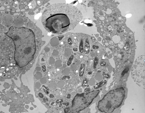
To replicate granuloma formation in an in vitro model, we infected human PBMCs with M. tuberculosis H37Ra or BCG and incubated for 5 days. At 24 h of incubation, PBMCs tended to form cellular aggregations of lymphocytes in the presence of H37Ra (Fig. 1A) or BCG (Fig. 1B). Corresponding control samples from the same donors cultured in the presence of Escherichia coli ATCC 25922 or Staphylococcus aureus ATCC 25923, or cultured in the absence of bacteria did not form these aggregates (Figs 1C, 1D, 1E) indicating that cellular aggregation forms specifically in response to M. tuberculosis infection. The granuloma-like shape of the cell aggregates formed following 24 h of M. tuberculosis H37Ra infection was confirmed by Wright-Giemsa staining (Fig. 1F). Transmission electron microscopy evidenced the engulfment of mycobacterial cells by phagocytes present in the cellular aggregates at 48 h post-infection (Fig. 2). The in vitro granulomatous structures persisted for 96 h, and then began to disappear.
Figure 1.
Infection of human PBMC with Mycobacterium tuberculosis resulted in formation of microscopic granulomas. PBMCs infected with: (A) H37Ra (100x), (B) BCG (100x), (C) Escherichia coli ATCC 25922 (200x), (D) Staphylococcus aureus ATCC 25923 (200x), or (E) uninfected PBMCs (100x). (F) Wright-Giemsa staining showing microgranulomas formed after 24 h of infection with H37Ra (400x).
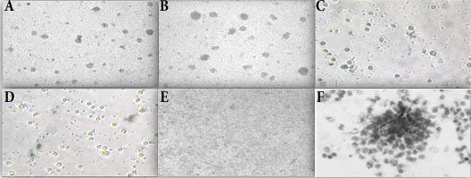
Figure 2.
Transmission electron microscopy of H37Ra-induced granuloma. After 48 hour post-infection, cellular aggregations were collected, fixed and embedded in an Epon-araldite resin. Sections of 0.5 µm were obtained, stained and observed under transmission electron microscope (4,000x). Multiple phagosomal vesicles containing H37Ra bacteria can be observed.
Microarray expression profiles of in vitro granulomas induced by H37Ra
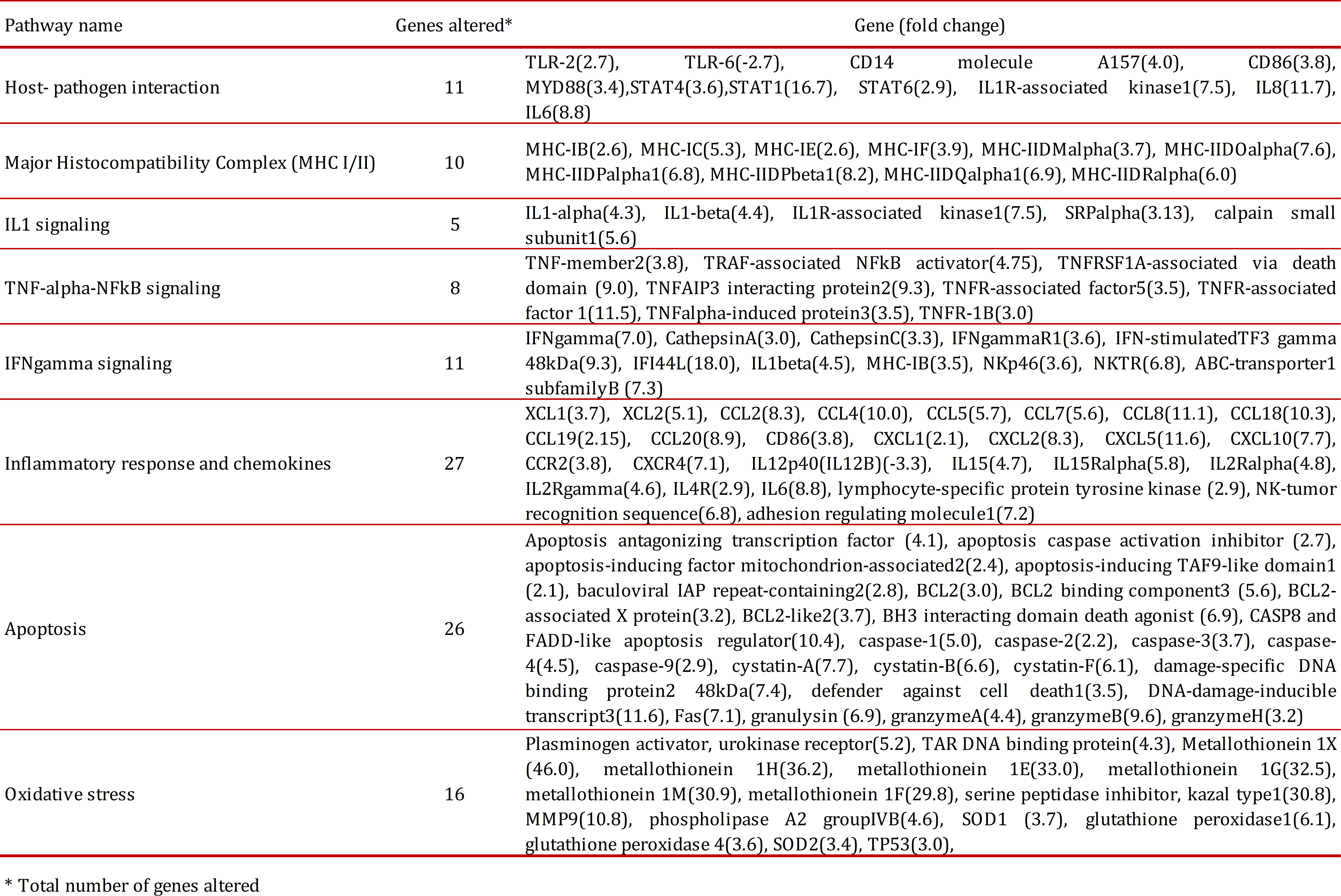
Of the total 25,690 genes analyzed with the microarrays, 2,195 were found overexpressed (fold change >-2, p <0.05) and 106 subexpressed (fold change <2, p< 0.05) in in vitro granulomas compared to untreated PBMCs. We found that 60% of altered genes were related to the immune response, such as antigenic processing, signaling pathways (TLR2, TNF, IL-6, IL-8, chemokines), 25% of genes were related to metabolic processes and 15% were related to oxidative stress and apoptosis (Table 1). We found overexpression of TLR2, CD14, CD86 and MyD88, which are main components of the TLR2 signaling of the innate immune response. We also found increased expression of both MHC-I and MHC-II molecules involved in antigen presentation by antigen-presenting cells (APC), a process essential to contain M. tuberculosis infection. In our in vitro granuloma model, at 24 h post-infection with M. tuberculosis H37Ra, we observed significant induction of a number of proinflammatory immune signaling pathways dependent on chemokines, including XCL1(3.7), XCL2(5.1), CCL2(8.3), CCL4(10.0), CCL5(5.7), CCL7(5.6), CCL8(11.1), CCL18(10.3), CCL19(2.15), CCL20(8.9), CXCL1(2.1), CXCL2(8.3), CXCL5(11.6), CXCL10(7.7), and chemokine receptors CCR2(3.8), and CXCR4(7.1). There was also increased expression of apoptosis related genes such as caspase-1 (5.0), caspase-2 (2.2), caspase-3 (3.7), caspase-4 (4.5) and caspase-9 (2.9). We also found increased expression of granulysin and granzymes A, B and H, main components of the cytoplasmic granules of cytotoxic T lymphocytes and natural killers, which are involved in cell-mediated apoptosis. We further observed increased expression of genes coding a type of peptidases involved in activation of CD8+ T cells, the cathepsins A, C, D, and W. We also identified altered expression of a number of chemokines not previously implicated in the immune response to M. tuberculosis, such as CCL8 (MCP-2), CCL7 (MCP-3), XCL1 (lymphotactin) and XCL2.
Real time PCR validation of gene expression for selected genes
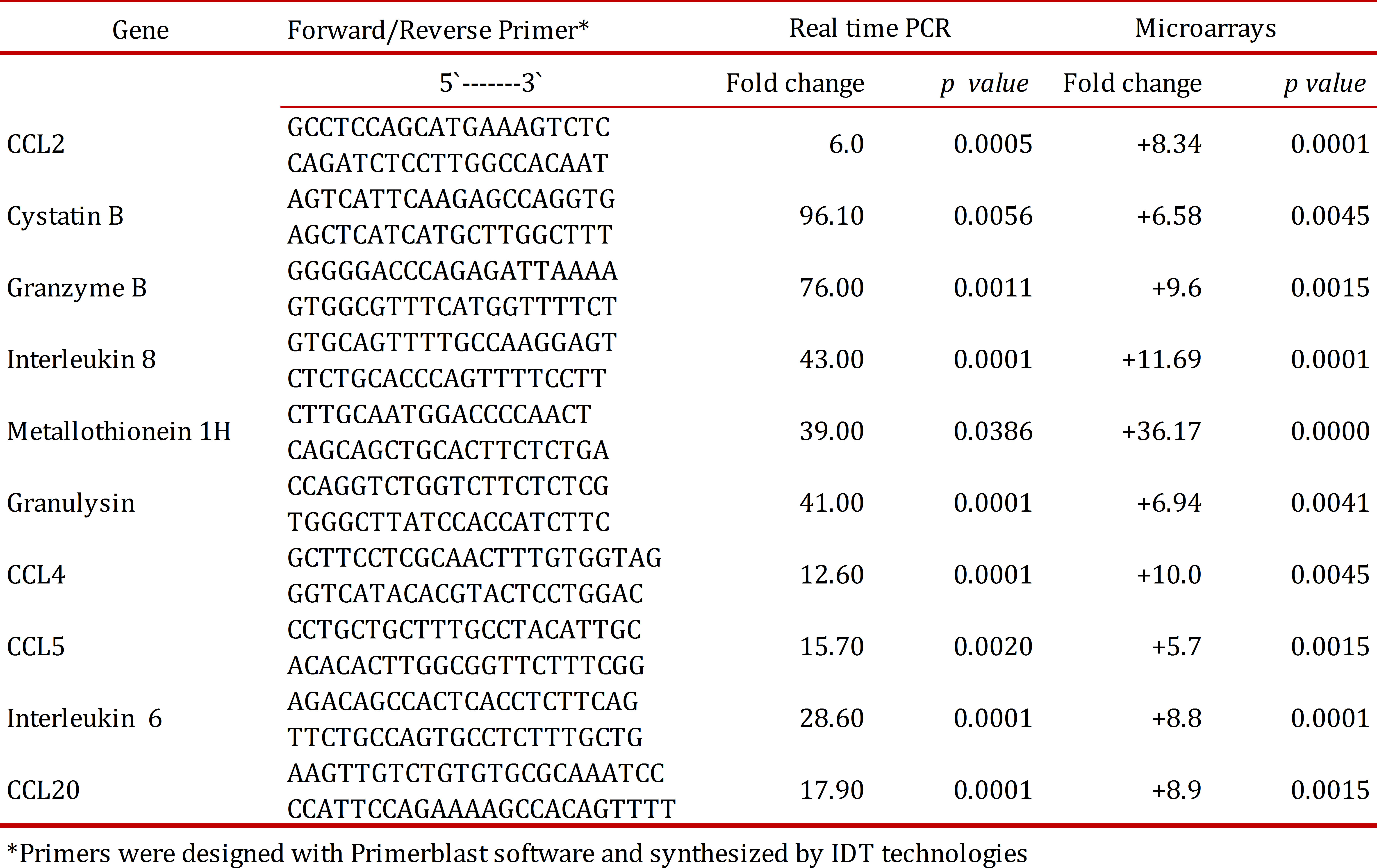
To validate microarray data, real time PCR quantitation was performed on a set of 10 genes including proinflammatory chemokines (CCL2, IL8, CCL5, CCL18), effector molecules involved in mechanisms of cell death dependent on cytotoxic T cells (cystatin B, granzyme B, granulysin), and metallothioneins (1H, 1M, 1G). Real time PCR results are presented in Table 2. Positive values indicate an increase in gene expression in in vitro granulomas at 24 h post-infection with H37Ra compared to uninfected PBMCs cultured for 24 h. Gene expression levels obtained by real time PCR were in agreement with corresponding levels observed in microarray experiments.
Discussion
We have used a previously described model for development of in vitro tuberculous granulomas to determine gene expression profiles associated to this process. Using this model, we observed that lymphocytes in human PBMCs clustered around infecting bacilli resembling micro-granuloma aggregates (Fig. 1). The micro-granulomas formed specifically in response to M. tuberculosis infection since these aggregates did not form in response to live E. coli or S. aureus nor did they form in uninfected samples. Development and maintenance of granulomas in the lung is the main host defense against M. tuberculosis 17,18. Microarray analysis of these in vitro granulomas allowed us to gain insights into early host-pathogen interactions taking place during granuloma formation in response to M. tuberculosis H37Ra (ATCC 25177). Initial recognition of M. tuberculosis by the innate immune response involves signaling through Toll-like receptor 2 (TLR2), which is influenced by several accessory receptors, mainly CD14 17,18. Lipomannans from mycobacterial species are agonists of TLR2, that, after ligand binding, induce macrophage activation characterized by cell surface expression of CD40 and CD86, cytokine and chemokine production, antigen presentation, among other innate immune responses 19. Macrophage activation is mediated through the adaptor protein myeloid differentiation factor 88 (MyD88), but independent of either TLR4 or TLR6 recognition 20,21. Consistent with these reports, in this study we found overexpression of TLR2, CD14, CD86 and MyD88, which supports the importance of TLR2 signaling in the innate immune response to M. tuberculosis H37Ra. The interactions between M. tuberculosis and innate immune cells result in secretion of chemokines and cytokines, of which IFN-γ and TNF are particularly important in TB. One important effect of IFN-γ is to activate macrophages and enhance their expression of MHC class II molecules, leading to increased antigen presentation to T cells, an event crucial for containment of mycobacterial infection 22. Here we found that H37Ra infection resulted in increased expression of both MHC-I and MHC-II molecules involved in antigen presentation by antigen-presenting cells (APC), a process essential to contain M. tuberculosis infection.
The most prominent effect observed in this study was the induction of many chemokine and cytokine genes. Several cytokines are known to play an important role in anti-TB immunity. Increased mRNA expression of IFN-γ TNF-α, IL-6, IL-8 and IL-12 in tuberculosis granuloma 23 and secretion of cytokines IL-1β, TNF-α and IL-6 in bronchoalveolar lavage fluid from involved sites of pulmonary TB have been reported 24. Transcriptome analysis of early granuloma lesions in the lungs of non-human primates exhibiting active TB, revealed a highly proinflammatory environment, expressing high levels of immune signaling pathways involving IFN-γ, TNF-α, JAK, STAT and CC/CXC chemokines 25. In our in vitro granuloma model, at 24 h post-infection with M. tuberculosis H37Ra, we observed significant induction of a number of proinflammatory immune signaling pathways dependent on chemokines, IFN-γ and TNF.
It has been widely shown that chemokines participate in the protective immune response to M. tuberculosis infection 26 and many of them are chemo-attractant for leucocytes to the site of infection. Our microarray analysis revealed significant differences in the expression of CXCL- and CCL- chemokines, such as CXCL1 (Gro-α), CXCL2 (Gro-β), CXCL5 (ENA-78), CXCL8 (IL-8) and CXCL10 (IP-10). Among them, CXCL5 transcripts were the most highly over-expressed. This chemokine, along with IL-8, is induced in response to proinflammatory cytokines such as IL-1 or TNF-α, which are potent neutrophil activators 27. Previous studies have shown that CXCL5 leads to an increase in the number of infiltrating neutrophils in bronchoalveolar carcinomas and infection by Rhinovirus 28; however, its role in tuberculosis infection is unclear 29.
We identified increased level of transcripts for CCL19 and CCL20 in our study. These two chemokines participate in the recruitment and activation of lymphocytes to sites of inflammation. The induction of numerous chemokines in early in vitro granuloma is an expected finding, since chemokine gradients are known to guide different immune effector cells to the site of infection. Chemokines have been demonstrated to be associated with mycobacterial infections, with appropriate levels of them required for preventing cells from migrating out of the granuloma, contributing in this way to maintain granuloma structure 30.
The role played by proinflammatory immune signaling pathways involving IFN-γ and TNF-α in protection against M. tuberculosis infection is well understood 31. Among the overexpressed genes found in this study we found IL15 and IL15R. Several studies have provided evidence that this cytokine pathway can enhance protective immune responses against M. tuberculosis infection 32. Other chemokine highly over-expressed in our in vitro granulomas was CXCL8 (IL-8). This chemokine is implicated in the formation of tuberculous granulomas and in immunity to M. tuberculosis 33.
Our study identified altered expression of different apoptosis related genes, including caspases, granulysin, granzymes, and cathepsins. It has been reported that M. tuberculosis infection causes apoptosis of neutrophils 34 and monocytes/macrophages 34,35. Macrophages are the primary host cells for M. tuberculosis and consequently, there is extensive cell death among this population of cells. Specifically, it has been shown that attenuated or avirulent strains of mycobacteria, such as BCG and H37Ra, primarily induce macrophage apoptosis, while virulent strains mainly induce necrosis. These observations have generated interest in the field of TB vaccine development since pro-apoptotic mycobacterial strains able to induce greater macrophage apoptosis may stimulate a quantitatively better T-cell response 36. Cathepsin C, found overexpressed in this study, participates in activation of granzymes and in the cell death processes mediated by cytolytic T cells 37,38, whereas cathepsin W associates with NK cells, thus playing an essential role in cytotoxicity 39. The increased expression of all these pro-apoptotic genes may reflect the attempt of the innate immune system to limit the infection by restricting the growth of M. tuberculosis, and reveals the role played by the various caspases, granzymes and cathepsins in this process.
There was also induction of metallothioneins and metalloproteases in the early granulomas analyzed in this study. Metallothioneins are low-molecular weight (6 to 8 KDa), cysteine-rich metal binding proteins which are induced by different stimuli, including oxidative stress, such as that generated during the respiratory burst by phagocytes 40. Metallothionein 1H has been implicated in induction of chemokines in vitro 41. There was a significant increase in the transcript levels of MMP9 and several cystatins. MMP9 is stabilized and protected by members of the cystatin superfamily without affecting its activity 42. Recent studies have shown the importance of MMP9 in interacting with M. tuberculosis secreted proteins and inducing the formation of granuloma lesions 43. It has been reported that mycobacterial lipomannan induces the expression of MMP9 in human macrophages through a mechanism dependent on TLR2 and CD14 44. The importance of Th1 cells in tuberculosis control is widely accepted 45, and our study clearly shows that M. tuberculosis H37Ra induces a strong Th1 response, reflected by the increased expression of Th1 cytokines such as IFN-γ, TNF-α, IL-6 and IL-1, that mediates successful resistance to M. tuberculosis infection 45,46. Our study supports this notion as we observed that many IFN-γ-inducible and regulated genes, such as IFI-16, IFI-30, IFI-44, IRF-3, IRF-7 y IRF-1 were increased.
Finally, in this study we identified altered expression of a number of chemokines that have not been previously implicated in the immune response to M. tuberculosis, such as CCL8 (MCP-2), CCL7 (MCP-3), XCL1 (lymphotactin) and XCL2. The potential role played by these chemokines in the establishment of the tuberculous granuloma warrants further investigation. All the changes described here took place in only 24 hours of infection of PBMCs by H37Ra M. tuberculosis strain, leading to early granuloma formation. Thus, this in vitro granuloma model using human PBMC is suitable for studying very early changes in gene expression taking place during induction of granuloma formation in response to M. tuberculosis infection. The model may be used to determine changes associated to different types of M. tuberculosis strains, including clinical isolates. It may also help in vaccine strategies, to decide which strains may be better candidates, based on the specific profiles of gene expression that they are able to induce.