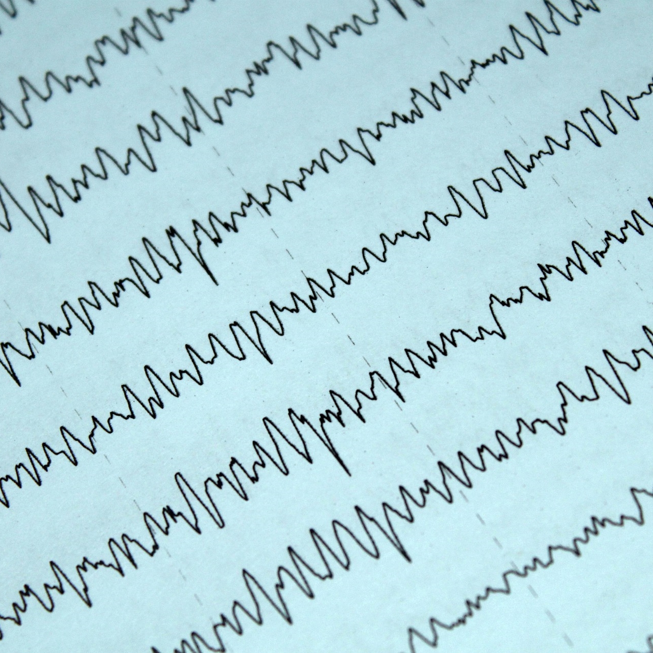
Triphasic waves and brain atrophy in patients with acute encephalopathy
Abstract
Introduction
Triphasic waves (TW) constitute an electroencephalographic pattern associated with certain kinds of encephalopathy. In addition, brain atrophy may be a predisposing factor linked with triphasic waves.
Objective
To compare the degree of brain atrophy and white matter disease between patients with acute encephalopathy with and without triphasic waves.
Methods
A retrospective observational study included adult patients with encephalopathy, with and without triphasic waves, hospitalized between 2016 and 2017. The degree of brain atrophy and white matter lesion were defined using the Global Cortical Atrophy and Age-Related White Matter Changes (ARWMC) scales, respectively. Scores were compared between groups. Mortality rates were registered.
Results
Sixteen patients with triphasic waves were identified matched by age and sex with 30 patients without triphasic waves. The mean age was 80 years in the triphasic waves group. Women represented 87.5%. Multifactorial encephalopathy was the most frequent diagnosis, followed by metabolic encephalopathy. Patients with triphasic waves had more brain atrophy (10.43 vs. 6.9, p= 0.03). Mean ARWMC was 9.43±6.5 and 8.5 ±7.89 in patients with and without triphasic waves, respectively (p= 0.5). The mortality rate was higher in the triphasic waves group (31.25 vs. 6.66% p= 0.02).
Conclusions
Patients with acute encephalopathy and triphasic waves had a higher degree of cerebral atrophy. This structural alteration may predispose to the appearance of triphasic waves. There was no significant difference in white matter lesion degree. The mortality of the triphasic waves group was high, so future studies are necessary to determine their prognostic value.
Authors
Downloads
Keywords
- Brain Atrophy
- Encephalopathy
- Triphasic waves
- Clinical Neurophysiology
- White matter
- electroencephalogram
References
Andraus MEC, Andraus CF, Alves-Leon SV. Periodic EEG patterns: importance of their recognition and clinical significance. Arq Neuropsiquiatr. 2012; 70(2): 145-151. https://doi.org/10.1590/S0004-282X2012000200014
Kaplan PW, Sutter R. Affair with triphasic waves - Thier striking presence, mysterious significance and cryptic origins: What are they? J Clin Neurophysiol, 2015; 32: 401-405. https://doi.org/10.1097/WNP.0000000000000151
Bermeo-Ovalle A. Triphasic Waves: swinging the pendulum back in this diagnostic dilemma. Epilepsy curr. 2017; 17(1): 40-42. https://doi.org/10.5698/1535-7511-17.1.40
Kaplan PW, Rossetti A. EEG Patterns and Imaging Correlations in Encephalopathy: Encephalopathy Part II. J Clin Neurophysiol. 2011. 28: 233-251. https://doi.org/10.1097/WNP.0b013e31821c33a0
American Psychiatric Association. Diagnostic and Statistical Manual of Mental Disorders. 5th ed. Washington, DC: American Psychiatric Association; 2013. https://doi.org/10.1176/appi.books.9780890425596
Hirsch LJ, LaRoche SM, Gaspard N, Gerrard E, Svoronos A, Herman ST et al. American Clinical Neurophysiology Society's Standardized Critical Care EEG Terminology: 2012 version. J Clin Neurophysiol, 2013; 30: 1-27. https://doi.org/10.1097/WNP.0b013e3182784729
Wahlund LO, Barkhof F, Fazekas F, Bronge L, Augustin M, Sjögren M, et al. A New Rating Scale for Age-Related White Matter Changes Applicable to MRI and CT. Stroke, 2001; 32: 1318-1322. https://doi.org/10.1161/01.STR.32.6.1318
Pasquier F, Leys D, Weerts JG, Mounier-Vehier F, Barkhof F, Scheltens P. Inter and Intraobserver Reproducibility of Cerebral Atrophy Assessment on MRI with Hemispheric Infarcts. Eur Neurol. 1996; 36: 268-272. https://doi.org/10.1159/000117270
Sutter R, Stevens RD, Kaplan PW. Clinical and imaging correlates of EEG patterns in hospitalized patients with encephalopathy. J Neurol, 2013; 260(4): 1087-1098. https://doi.org/10.1007/s00415-012-6766-1
Sutter R, Stevens RD, Kaplan, PW. Significance of triphasic waves in patients with acute encephalopathy: A nine-year cohort study. Clin Neurophysiol. 2013; 124: 1952-1958. https://doi.org/10.1016/j.clinph.2013.03.031
Sutter R, Kaplan PW. Uncovering clinical and radiological associations of triphasic waves in acute encephalopathy: a case-control study. Eur J Neurol, 2014; 21: 660-666. https://doi.org/10.1111/ene.12372
van-Putten MJAM, Hofmeijjer J. Generalized periodic discharges: pathophysiology and clinical considerations. Epilepsy Behav. 2015; 49: 228- 233. https://doi.org/10.1016/j.yebeh.2015.04.007
Sully K, Husain AM. Generalized Periodic Discharges: A topical Review. J Clin Neurophysiol. 2018; 35: 199-207 https://doi.org/10.1097/WNP.0000000000000460
Fjell AM, McEvoy L, Holland D, Dale AM, Walhovd KB.What is normal in normal aging? Effects of Aging, Amyloid and Alzheimer's Disease on the Cerebral Cortex and the Hippocampus. Prog Neurobiol. 2014; 117: 20-40. https://doi.org/10.1016/j.pneurobio.2014.02.004
Festari C, Ferrari C, Nicolosi V, Rossi R, Altomare D, Galluzzi S, et al. Age-related white matter changes scale: normative data on 1,439 persons. Alzheimers Dement. 2017; 13(7):P443. https://doi.org/10.1016/j.jalz.2017.06.443

Copyright (c) 2022 Universidad del Valle

This work is licensed under a Creative Commons Attribution-NonCommercial 4.0 International License.
The copy rights of the articles published in Colombia Médica belong to the Universidad del Valle. The contents of the articles that appear in the Journal are exclusively the responsibility of the authors and do not necessarily reflect the opinions of the Editorial Committee of the Journal. It is allowed to reproduce the material published in Colombia Médica without prior authorization for non-commercial use

 https://orcid.org/0000-0001-7645-084X


