
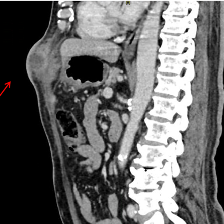
Abdominal actinomycosis by Actinomyces shaaliae georgiae mimicking neoplasia Case report

Article Sidebar
Main Article Content
Objective
This study aimed to investigate whether serum Interleukin 36 gamma (IL-36γ) levels in pediatric patients with allergic rhinitis correlate with disease severity (mild, moderate, severe) and duration (intermittent, persistent). Additionally, we assessed the potential of IL-36γ as a biomarker and its role in disease pathogenesis to inform future treatment strategies.
Methods
In this cross-sectional observational study, pediatric patients with allergic rhinitis from outpatient clinics were compared with healthy controls. Serum IL-36γ levels were measured from blood samples and analyzed across subgroups based on disease severity and duration.
Results
Fifty patients with allergic rhinitis and forty controls were included. IL-36γ levels were higher in the patient group, with borderline significance (p = 0.050). Female patients had significantly higher IL-36γ levels than male controls (p= 0.044).
Conclusion
This is the first study to evaluate IL-36γ levels in pediatric allergic rhinitis. Although the difference between groups showed borderline significance, larger studies may confirm these findings. The observed gender-related difference suggests IL-36γ could be a potential biomarker. Additionally, a significant negative correlation with total IgE and a nonsignificant negative correlation with eosinophil counts were noted.
- Actinomyces
- Actinomycosis
- Abdomen
- Actinomycetaceae
- Actinomyces georgiae
Mateo Londoño Barrientos, Universidad Escuela de Ingeniería de Antioquia, Envigado, Colombia
 https://orcid.org/0009-0001-8524-9512
Universidad Escuela de Ingeniería de Antioquia, Envigado, Colombia
Carlos Alberto Lopez Zapata, Hospital Pablo Tobón Uribe, Medellín, Colombia
https://orcid.org/0000-0001-9682-7835
Hospital Pablo Tobón Uribe, Medellín, Colombia
Laura Álvarez Herrera, Hospital Pablo Tobón Uribe, Medellín, Colombia
https://orcid.org/0009-0003-4169-8724
1 Universidad Escuela de Ingeniería de Antioquia, Envigado, Colombia
2 Hospital Pablo Tobón Uribe, Medellín, Colombia
Marco Frusteri, Universidad Escuela de Ingeniería de Antioquia, Envigado, Colombia
https://orcid.org/0009-0004-5222-7757
1 Universidad Escuela de Ingeniería de Antioquia, Envigado, Colombia
2 Hospital Pablo Tobón Uribe, Medellín, Colombia
Carlos Andres Delgado Lopez, Hospital Pablo Tobón Uribe, Medellín, Colombia
https://orcid.org/0000-0002-0341-9793
Hospital Pablo Tobón Uribe, Medellín, Colombia
Wong VK, Turmezei TD, Weston VC: Actinomycosis. BMJ. 2011, 343:6099. https://doi.org/10.1136/bmj.d6099 PMid:21990282 DOI: https://doi.org/10.1136/bmj.d6099
Valour F, Sénéchal A, Dupieux C, et al.: Actinomycosis: etiology, clinical features, diagnosis, treatment, and management. Infect Drug Resist. 2014, 7:183-97. https://doi.org/10.2147/IDR.S39601 PMid:25045274 PMCid:PMC4094581 DOI: https://doi.org/10.2147/IDR.S39601
Könönen E, Wade WG: Actinomyces and related organisms in human infections. Clin Microbiol Rev. 2015, 28:419-42. https://doi.org/10.1128/CMR.00100-14 PMid:25788515 PMCid:PMC4402957 DOI: https://doi.org/10.1128/CMR.00100-14
Lewis RP, Sutter VL, Finegold SM: Bone infections involving anaerobic bacteria. Medicine (Baltimore). 1978, 57:279-305. https://doi.org/10.1097/00005792-197807000-00001 PMid:207946 DOI: https://doi.org/10.1097/00005792-197807000-00001
Mabeza GF, Macfarlane J: Pulmonary actinomycosis. Eur Respir J. 2003, 21:545-51. https://doi.org/10.1183/09031936.03.00089103 PMid:12662015 DOI: https://doi.org/10.1183/09031936.03.00089103
Bennett JE, Dolin R, Blaser MJ. Mandell, Douglas, and Bennett's principles and practice of infectious diseases E-Book: 2-volume set. Elsevier health sciences, 2019.
Heo SH, Shin SS, Kim JW, et al.: Imaging of actinomycosis in various organs: a comprehensive review. Radiographics. 2014, 34:19-33. https://doi.org/10.1148/rg.341135077 PMid:24428279 DOI: https://doi.org/10.1148/rg.341135077
Triantopoulou C, der Molen AV, Es ACV, Giannila M: Abdominopelvic actinomycosis: spectrum of imaging findings and common mimickers. Acta Radiol Short Rep. 2014, 3:2047981614524570. https://doi.org/10.1177/2047981614524570 DOI: https://doi.org/10.1177/2047981614524570
PMid:24778807 PMCid:PMC4001438
Sung HY, Lee IS, Kim SI, et al.: Clinical Features of Abdominal Actinomycosis: A 15-year Experience of A Single Institute. J Korean Med Sci. 2011, 26:932-7. https://doi.org/10.3346/jkms.2011.26.7.932 PMid:21738348 PMCid:PMC3124725 DOI: https://doi.org/10.3346/jkms.2011.26.7.932
Rahimkhani M, Mordadi A, Kazemian K, Khalili H: Comparison of helicobacter pylori detection methods: It's association with leukocytosis and monocytosis. Infectious Disorders - Drug Targets. 2020, 20(6):920-924. https://doi.org/10.2174/1871526520666200707113955 PMid:32634084 DOI: https://doi.org/10.2174/1871526520666200707113955
Choi M-M, Baek JH, Lee JN, Park S, Lee W-S: Clinical features of abdominopelvic actinomycosis: report of twenty cases and literature review. Yonsei Med J. 2009, 50:555-9. https://doi.org/10.3349/ymj.2009.50.4.555 PMid:19718405 PMCid:PMC2730619 DOI: https://doi.org/10.3349/ymj.2009.50.4.555
Vasilescu AM, Târcoveanu E, Lupascu C, Blaj M, Lupascu Ursulescu C, Bradea C: Abdominopelvic Actinomycosis-The Diagnostic and Therapeutic Challenge of the Most Misdiagnosed Disease. Life. 2022, 12:447. https://doi.org/10.3390/life12030447 PMid:35330198 PMCid:PMC8954618 DOI: https://doi.org/10.3390/life12030447
Arslan RS, Koca YS, Beyoğlu R, Yenipazar AE: Appendecular actinomycosis: A case series of 14 patients. Med Clin (Barc). 2024, 162:500-4. https://doi.org/10.1016/j.medcli.2024.02.005 PMid:38570296 DOI: https://doi.org/10.1016/j.medcli.2024.02.005
Downloads


Accepted 2025-09-30
Published 2025-06-30

This work is licensed under a Creative Commons Attribution-NonCommercial 4.0 International License.
The copy rights of the articles published in Colombia Médica belong to the Universidad del Valle. The contents of the articles that appear in the Journal are exclusively the responsibility of the authors and do not necessarily reflect the opinions of the Editorial Committee of the Journal. It is allowed to reproduce the material published in Colombia Médica without prior authorization for non-commercial use