
Morphometric analysis and tortuosity typing of the large intestine segments on computed tomography colonography with artificial intelligence Morfometría y tipificación de tortuosidad del colon

Keywords:
Computed tomography, large intestine, colonography, morphometry, tortuosity, artificial intelligenceArticle Sidebar
Accepted 2024-08-15
Published 2024-11-27
Main Article Content
Background:
Morphological properties such as length and tortuosity of the large intestine segments play important roles, especially in interventional procedures like colonoscopy.
Objective:
Using computed tomography (CT) colonoscopy images, this study aimed to examine the morphological features of the colon's anatomical sections and investigate the relationship of these sections with each other or with age groups. The shapes of the transverse colon were analyzed using artificial intelligence.
Materials and Methods:
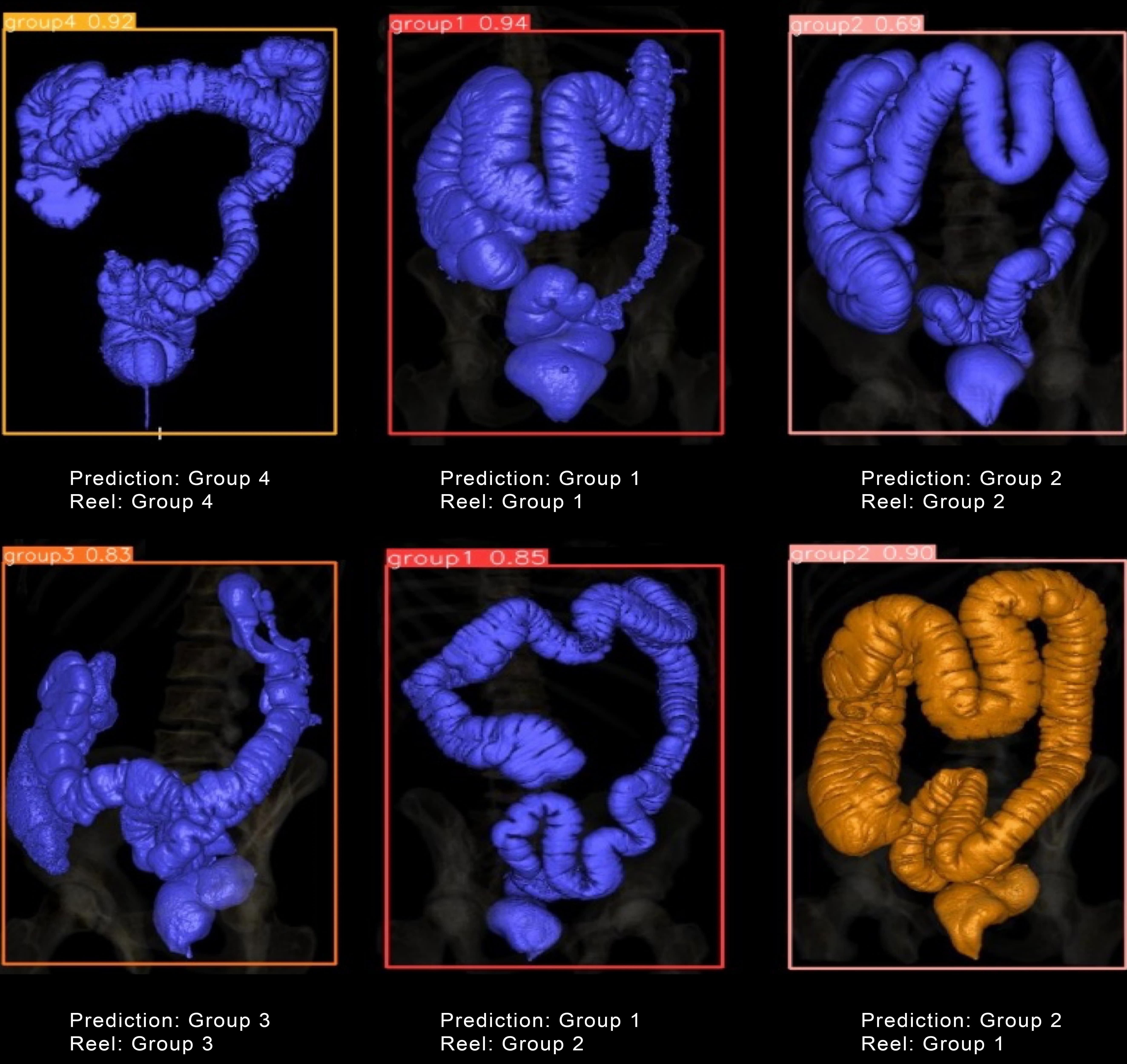
The study was conducted as a two- and three-dimensional examination of CT colonography images of people between 40 and 80 years old, which were obtained retrospectively. An artificial intelligence algorithm (YOLOv8) was used for shape detection on 3D colon images.
Results:
160 people with a mean age of 89 men and 71 women included in the study was 57.79±8.55 and 56.55±6.60, respectively, and there was no statistically significant difference (p=0.24). The total colon length was 166.11±25.07 cm for men and 158.73±21.92 cm for women, with no significant difference between groups (p=0.12). As a result of the training of the model Precision, Recall, and mAP were found to be 0.8578, 0.7940, and 0.9142, respectively.
Conclusion:
The study highlights the importance of understanding the type and morphology of the large intestine for accurate interpretation of CT colonography results and effective clinical management of patients with suspected large intestine abnormalities. Furthermore, this study showed that 88.57% of the images in the test data set were detected correctly and that AI can play an important role in colon typing.
Hadi Sasani, Tekirdag Namik Kemal University, Faculty of Medicine, Department of Radiology, Tekirdag, Turkey.
 https://orcid.org/0000-0001-6236-4123
Mazhar Ozkan, Tekirdag Namık Kemal University, School of Medicine, Department of Anatomy, Tekirdag, Turkey
https://orcid.org/0000-0002-8745-2493
Mehmet Ali Simsek, Tekirdag Namik Kemal University
https://orcid.org/0000-0002-6127-2195
Mahmut Sasani, Bezmi Alem Vakif University, Faculty of Medicine, Istanbul, Turkey
https://orcid.org/0000-0002-2313-1754
Standring S. Gray's Anatomy e-Book: The Anatomical Basis of Clinical Practice. 41st ed. Elsevier Health Sciences; 2016.
Faiss S. The missed colorectal cancer problem. Dig Dis Basel Switz. 2011; 29 Suppl 1: 60-63; https://doi.org/10.1159/000331119
Petryszyn PW, Kempiński R, Michałowicz J, et al. Non-medical costs of colonoscopy. Przeglad Gastroenterol. 2014; 9(5): 270-4; https://doi.org/10.5114/pg.2014.46161
Scheirey CD, Fowler KJ, Therrien JA, et al. ACR Appropriateness Criteria® Acute Nonlocalized Abdominal Pain. J Am Coll Radiol. 2018; 15(11, Supplement): S217-S231; https://doi.org/10.1016/j.jacr.2018.09.010
Malik J, Kiranyaz S, Kunhoth S, et al. Colorectal Cancer Diagnosis from Histology Images: A Comparative Study. 2019; doi: 10.48550/arXiv.1903.11210.
Gupta P, Chiang S-F, Sahoo PK, et al. Prediction of Colon Cancer Stages and Survival Period with Machine Learning Approach. Cancers. 2019; 11(12): 2007; https://doi.org/10.3390/cancers11122007
Ogony J, de Bel T, Radisky DC, et al. Towards defining morphologic parameters of normal parous and nulliparous breast tissues by artificial intelligence. Breast Cancer Res BCR. 2022; 24(1): 45. https://doi.org/10.1186/s13058-022-01541-z
Smith K, Clark K, Bennett W, et al. Data From CT COLONOGRAPHY. 2015; doi: https://doi.org/10.7937/K9/TCIA.2015.NWTESAY1.
Redmon J, Divvala S, Girshick R, et al. You Only Look Once: Unified, Real-Time Object Detection. In: 2016 IEEE Conference on Computer Vision and Pattern Recognition (CVPR). 2016; pp. 779-88; https://doi.org/10.1109/CVPR.2016.91
Su Y, Cheng B, Cai Y. Detection and Recognition of Traditional Chinese Medicine Slice Based on YOLOv8. In: 2023 IEEE 6th International Conference on Electronic Information and Communication Technology (ICEICT). 2023; pp. 214-7; https://doi.org/10.1109/ICEICT57916.2023.10245026
Xiao B, Nguyen M, Yan WQ. Fruit ripeness identification using YOLOv8 model. Multimed Tools Appl. 2023; doi: 10.1007/s11042-023-16570-9.
https://doi.org/10.1007/s11042-023-16570-9
Atkin W, Dadswell E, Wooldrage K, et al. Computed tomographic colonography versus colonoscopy for investigation of patients with symptoms suggestive of colorectal cancer (SIGGAR): a multicentre randomised trial. The Lancet. 2013; 381(9873): 1194-202; https://doi.org/10.1016/S0140-6736(12)62186-2
Ganeshan D, Elsayes KM, Vining D. Virtual colonoscopy: Utility, impact and overview. World J Radiol. 2013; 5(3): 61-7; https://doi.org/10.4329/wjr.v5.i3.61
Abe T, Ujiie A, Taguchi Y, et al. Anomalous inferior mesenteric artery supplying the ascending, transverse, descending, and sigmoid colons. Anat Sci Int. 2018; 93(1): 144-8; https://doi.org/10.1007/s12565-017-0401-2
Wozniak S, Pawlus A, Grzelak J, et al. Acute colonic flexures: the basis for developing an artificial intelligence-based tool for predicting the course of colonoscopy. Anat Sci Int. 2023; 98(1): 136-42; https://doi.org/10.1007/s12565-022-00681-8
Khashab MA, Pickhardt PJ, Kim DH, et al. Colorectal anatomy in adults at computed tomography colonography: normal distribution and the effect of age, sex, and body mass index. Endoscopy. 2009; 41(8): 674-8; https://doi.org/10.1055/s-0029-1214899
Sadahiro S, Ohmura T, Yamada Y, et al. Analysis of length and surface area of each segment of the large intestine according to age, sex and physique. Surg Radiol Anat SRA. 1992; 14(3): 251-7; https://doi.org/10.1007/BF01794949
Utano K, Nagata K, Honda T, et al. Bowel habits and gender correlate with colon length measured by CT colonography. Jpn J Radiol. 2022; 40(3): 298-307; https://doi.org/10.1007/s11604-021-01204-7
Müller-Lissner SA, Kamm MA, Scarpignato C, et al. Myths and misconceptions about chronic constipation. Am J Gastroenterol. 2005; 100(1): 232-42; https://doi.org/10.1111/j.1572-0241.2005.40885.x
World Health Organization. International Agency for Research on Cancer. 2023.
Coffey JC. Surgical anatomy and anatomic surgery - Clinical and scientific mutualism. The Surgeon. 2013; 11(4): 177-82; https://doi.org/10.1016/j.surge.2013.03.002
Tirkes T, Sandrasegaran K, Patel AA, et al. Peritoneal and retroperitoneal anatomy and its relevance for cross-sectional imaging. Radiogr Rev Publ Radiol Soc N Am Inc. 2012; 32(2): 437-51; https://doi.org/10.1148/rg.322115032
Alazmani A, Hood A, Jayne D, et al. Quantitative assessment of colorectal morphology: Implications for robotic colonoscopy. Med Eng Phys. 2016; 38(2): 148-54; https://doi.org/10.1016/j.medengphy.2015.11.018
Primal Pictures. Anatomical Variations: The Transverse Colon. n.d. Available from: https://www.primalpictures.com/blogs/transverse-colon-anatomical-variation/.
Downloads

This work is licensed under a Creative Commons Attribution-NonCommercial 4.0 International License.
The copy rights of the articles published in Colombia Médica belong to the Universidad del Valle. The contents of the articles that appear in the Journal are exclusively the responsibility of the authors and do not necessarily reflect the opinions of the Editorial Committee of the Journal. It is allowed to reproduce the material published in Colombia Médica without prior authorization for non-commercial use