Variability between Clarke's angle and Chippaux-Smirak index for the diagnosis of flat feet
Keywords:
Footprint, pedigraph, chippaux-smirak index, clarke's angleMain Article Content
Background
The measurements used in diagnosing biomechanical pathologies vary greatly. The aim of this study was to determine the concordance between Clarke’s angle and Chippaux-Smirak index, and to determine the validity of Clarke’s angle using the Chippaux-Smirak index as a reference.
Methods
Observational study in a random population sample (n=1002) in A Coruña (Spain). After informed patient consent and ethical review approval, a study was conducted of anthropometric variables, Charlson comorbidity score, and podiatric examination. Descriptive analysis and multivariate logistic regression were performed.
Results
The prevalence of flat feet, using a podoscope, was 19.0% for the left foot and 18.9% for the right foot, increasing with age.
The prevalence of flat feet according to the Chippaux-Smirak index or Clarke’s angle increases significantly, reaching 62.0% and 29.7% respectively.
The concordance (kappa I) between the indices according to age groups varied between 0.25–0.33 (left foot) and 0.21–0.30 (right foot). The intraclass correlation coefficient (ICC) between the Chippaux-Smirak index and Clarke’s angle was -0.445 (left foot) and -0.424 (right foot).
After adjusting for age, body mass index (BMI), comorbidity score and gender, the only variable with an independent effect to predict discordance was the BMI (OR=0.969; 95% CI:0.94-0.998).
Conclusion
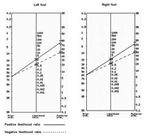
There is little concordance between the indices studied for the purpose of diagnosing foot arch pathologies. In turn, Clarke’s angle has a limited sensitivity in diagnosing flat feet, using the Chippaux-Smirak index as a reference. This discordance decreases with higher BMI values.
Hajjaj FM, Salek MS, Basra MK, Finlay AY: Non-clinical influences on clinical decision-making: a major challenge to evidence-based practice. J R Soc Med 2010, 103(5):178-187. DOI: https://doi.org/10.1258/jrsm.2010.100104
Rome K, Ashford RL, Evans A: Non-surgical interventions for paediatric pes planus. Cochrane Database Syst Rev 2010(7):CD006311. DOI: https://doi.org/10.1002/14651858.CD006311.pub2
Schwartz L, Britten RH, Thompson LR: Studies in Physical Development and Posture. I. The Effect of Exercise on the Physical Condition and Development of Adolescent Boys. Public Health Bulletin 1928(179).
Shiang TY, Lee SH, Lee SJ, Chu WC: Evaluating different footprint parameters as a predictor of arch height. IEEE Eng Med Biol Mag 1998, 17(6):62-66. DOI: https://doi.org/10.1109/51.731323
Xiong S, Goonetilleke RS, Witana CP, Weerasinghe TW, Au EY: Foot arch characterization: a review, a new metric, and a comparison. J Am Podiatr Med Assoc 2010, 100(1):14-24. DOI: https://doi.org/10.7547/1000014
Chen KC, Yeh CJ, Kuo JF, Hsieh CL, Yang SF, Wang CH: Footprint analysis of flatfoot in preschool-aged children. Eur J Pediatr 2011, 170(5):611-617. DOI: https://doi.org/10.1007/s00431-010-1330-4
Evans AM: Screening for foot problems in children: is this practice justifiable? J Foot Ankle Res 2012, 5(1):18. DOI: https://doi.org/10.1186/1757-1146-5-18
Chen KC, Tung LC, Yeh CJ, Yang JF, Kuo JF, Wang CH: Change in flatfoot of preschool-aged children: a 1-year follow-up study. Eur J Pediatr 2013, 172(2):255-260. DOI: https://doi.org/10.1007/s00431-012-1884-4
Pita-Fernández S, González-Martín C, Seoane-Pillado T, López-Calviño B, Pértega-Díaz S, Gil-Guillén V: Validity of footprint analysis to determine flatfoot using clinical diagnosis as the gold standard in a random sample aged 40 years and older. J Epidemiol 2015, 25(2):148-154. DOI: https://doi.org/10.2188/jea.JE20140082
Charlson ME, Pompei P, Ales KL, MacKenzie CR: A new method of classifying prognostic comorbidity in longitudinal studies: development and validation. J Chronic Dis 1987, 40(5):373-383. DOI: https://doi.org/10.1016/0021-9681(87)90171-8
Queen RM, Mall NA, Hardaker WM, Nunley JA: Describing the medial longitudinal arch using footprint indices and a clinical grading system. Foot Ankle Int 2007, 28(4):456-462. DOI: https://doi.org/10.3113/FAI.2007.0456
Mathieson I, Upton D, Prior TD: Examining the validity of selected measures of foot type: a preliminary study. J Am Podiatr Med Assoc 2004, 94(3):275-281. DOI: https://doi.org/10.7547/0940275
Wrobel JS, Armstrong DG: Reliability and validity of current physical examination techniques of the foot and ankle. J Am Podiatr Med Assoc 2008, 98(3):197-206. DOI: https://doi.org/10.7547/0980197
Fascione JM, Crews RT, Wrobel JS: Dynamic footprint measurement collection technique and intrarater reliability: ink mat, paper pedography, and electronic pedography. J Am Podiatr Med Assoc 2012, 102(2):130-138. DOI: https://doi.org/10.7547/1020130
Young CC, Niedfeldt MW, Morris GA, Eerkes KJ: Clinical examination of the foot and ankle. Prim Care 2005, 32(1):105-132. DOI: https://doi.org/10.1016/j.pop.2004.11.002
Akobeng AK: Understanding diagnostic tests 2: likelihood ratios, pre- and post-test probabilities and their use in clinical practice. Acta Paediatr 2007, 96(4):487-491. DOI: https://doi.org/10.1111/j.1651-2227.2006.00179.x
Aranceta J, Pérez Rodrigo C, Serra Majem L, Ribas L, Quiles Izquierdo J, Vioque J, Foz M: [Prevalence of obesity in Spain: the SEEDO'97 study. Spanish Collaborative Group for the Study of Obesity]. Med Clin (Barc) 1998, 111(12):441-445.
Mokdad AH, Ford ES, Bowman BA, Dietz WH, Vinicor F, Bales VS, Marks JS: Prevalence of obesity, diabetes, and obesity-related health risk factors, 2001. JAMA 2003, 289(1):76-79. DOI: https://doi.org/10.1001/jama.289.1.76
Dunn JE, Link CL, Felson DT, Crincoli MG, Keysor JJ, McKinlay JB: Prevalence of foot and ankle conditions in a multiethnic community sample of older adults. Am J Epidemiol 2004, 159(5):491-498. DOI: https://doi.org/10.1093/aje/kwh071
Nguyen US, Hillstrom HJ, Li W, Dufour AB, Kiel DP, Procter-Gray E, Gagnon MM, Hannan MT: Factors associated with hallux valgus in a population-based study of older women and men: the MOBILIZE Boston Study. Osteoarthritis Cartilage 2010, 18(1):41-46. DOI: https://doi.org/10.1016/j.joca.2009.07.008
Robbins JM: Recognizing, treating, and preventing common foot problems. Cleve Clin J Med 2000, 67(1):45-47, 51-42, 55-46. DOI: https://doi.org/10.3949/ccjm.67.1.45
Shibuya N, Jupiter DC, Ciliberti LJ, VanBuren V, La Fontaine J: Characteristics of adult flatfoot in the United States. J Foot Ankle Surg 2010, 49(4):363-368. DOI: https://doi.org/10.1053/j.jfas.2010.04.001
Atamturk D: [Relationship of flatfoot and high arch with main anthropometric variables]. Acta Orthop Traumatol Turc 2009, 43(3):254-259. DOI: https://doi.org/10.3944/AOTT.2009.254
Evans AM, Rome K: A Cochrane review of the evidence for non-surgical interventions for flexible pediatric flat feet. Eur J Phys Rehabil Med 2011, 47(1):69-89.
Pfeiffer M, Kotz R, Ledl T, Hauser G, Sluga M: Prevalence of flat foot in preschool-aged children. Pediatrics 2006, 118(2):634-639. DOI: https://doi.org/10.1542/peds.2005-2126
García-Rodríguez A, Martín-Jiménez F, Carnero-Varo M, Gómez-Gracia E, Gómez-Aracena J, Fernández-Crehuet J: Flexible flat feet in children: a real problem? Pediatrics 1999, 103(6):e84. DOI: https://doi.org/10.1542/peds.103.6.e84
Papuga MO, Burke JR: The reliability of the Associate Platinum digital foot scanner in measuring previously developed footprint characteristics: a technical note. J Manipulative Physiol Ther 2011, 34(2):114-118. DOI: https://doi.org/10.1016/j.jmpt.2010.12.008
Downloads

This work is licensed under a Creative Commons Attribution-NonCommercial 4.0 International License.
The copy rights of the articles published in Colombia Médica belong to the Universidad del Valle. The contents of the articles that appear in the Journal are exclusively the responsibility of the authors and do not necessarily reflect the opinions of the Editorial Committee of the Journal. It is allowed to reproduce the material published in Colombia Médica without prior authorization for non-commercial use

