New clues to understand gastroschisis. Embryology, pathogenesis and epidemiology
Abstract
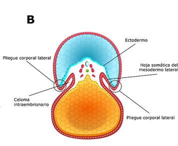
Gastroschisis is a congenital structural defect of the abdominal wall, most often to the right of the umbilicus, through which the abdominal viscera protrude. For a long time, its developmental, etiological, and epidemiological aspects have been a hot topic of controversy. However, recent findings suggest the involving of genetic and chromosomal alterations and the existence of a stress-inducing pathogenetic pathway, in which risk factors such as demographic and environmental ones can converge.
To expand the frontier of knowledge about a malformation that has shown a growing global prevalence, we have conducted a review of the medical literature that gathers information on the embryonic development of the ventral body wall, the primitive intestine and the ring-umbilical cord complex, as well as on the theories about its origin, pathogenesis and recent epidemiological evidence, for which we consulted bibliographic databases and standard search engines.
Authors
Downloads
Keywords
- Gastroschisis
- abdominal abnormalities
- Vitelline Duct
- Ectopia Cordis
- Genetic Predisposition to Disease
References
Calder J. Two examples of children with preternatural conformation of the guts. Medical Essays and observations, Vol I. T, W Ruddimans, Medical Society of Edinburgh. 1733; 1:203
Curry JI, McKinney P, Thornton JG, Stringer MD. The aetiology of gastroschisis. BJOG. 2000; 107(11): 1339-1346. https://doi.org/10.1111/j.1471-0528.2000.tb11645.x
Torres US, Portela-Oliveira E, Braga F del C, Werner H Jr, Daltro PA, Souza AS. When closure fails: what the radiologist needs to know about the embryology, anatomy, and prenatal imaging of ventral body wall defects. Semin Ultrasound CT MR. 2015; 36(6): 522-536. https://doi.org/10.1053/j.sult.2015.01.001
Chesley PM, Ledbetter DJ, Meehan JJ, Oron AP, Javid PJ. Contemporary trends in the use of primary repair for gastroschisis in surgical infants. Am J Surg. 2015; 209(5): 901-905. https://doi.org/10.1016/j.amjsurg.2015.01.012
Bhat V, Moront M, Bhandari V. Gastroschisis: A State-of-the-Art Review. Children (Basel). 2020; 7(12):302. https://doi.org/10.3390/children7120302
Mandelia A, Agarwala S, Sharma N, Solanki S, Panda S. Left-sided Gastroschisis: A rare congenital anomaly. J Clin Diagn Res. 2013; 7(10): 2300-2302. https://doi.org/10.7860/JCDR/2013/6161.3505
Schierz IAM, Pinello G, Giuffrè M, Corsello G. An unusual association of left-sided gastroschisis and persistent right umbilical vein. Clin Case Rep. 2018; 6(12): 2511-2512. https://doi.org/10.1002/ccr3.1897
Feldkamp ML, Carey JC, Sadler TW. Development of gastroschisis: review of hypotheses, a novel hypothesis, and implications for research. Am J Med Genet Part A. 2007; 639-652. https://doi.org/10.1002/ajmg.a.31578
Sadler TW. The embryologic origin of ventral body wall defects. Semin Pediatr Surg. 2010; 19(3): 209-214. https://doi.org/10.1053/j.sempedsurg.2010.03.006
Stevenson RE, Rogers RC, Chandler JC, Gauderer MW, Hunter AG. Escape of the yolk sac: a hypothesis to explain the embryogenesis of gastroschisis. Clin Genet. 2009; 75(4): 326-333. https://doi.org/10.1111/j.1399-0004.2008.01142.x
Melov SJ, Tsang I, Cohen R, Badawi N, Walker K, Soundappan SSV et al. Complexity of gastroschisis predicts outcome: epidemiology and experience in an Australian tertiary centre. BMC Pregnancy Childbirth. 2018; 18(1): 1-9. https://doi.org/10.1186/s12884-018-1867-1
Salinas-Torres VM, Salinas-Torres RA, Cerda-Flores RM, Martínez-de-Villarreal LE. Familial occurrence of gastroschisis: a population-based overview on recurrence risk, sex-dependent influence, and geographical distribution. Pediatr Surg Int. 2018; 34(3): 277-282. https://doi.org/10.1007/s00383-018-4224-6
Salinas-Torres VM, Gallardo-Blanco HL, Salinas-Torres RA, Cerda-Flores RM, Lugo-Trampe JJ, Villarreal-Martínez DZ et al. Bioinformatic Analysis of gene variants from gastroschisis recurrence identifies multiple novel pathogenetic pathways: implication for the closure of the ventral body wall. Int J Mol Sci. 2019; 20(9): 2295. https://doi.org/10.3390/ijms20092295
Feldkamp ML, Krikov S, Gardner J, Madsen MJ, Darlington T, Sargent R et al. Shared genomic segments in high‐risk multigenerational pedigrees with gastroschisis. Birth Defects Res. 2019; 111(20): 1655-1664. https://doi.org/10.1002/bdr2.1567
Sadler TW. Langman Embriología Médica, 14ª edición. Philadelphia: Wolters Kluwer. 2019. p 84, p 97-100
Sadler TW, Feldkamp ML. The embryology of body wall closure: Relevance to gastroschisis and other ventral body wall defects. Am J Med Genetics Part C (Seminars in Medical Genetics). 2008; 148C (3): 180-185. https://doi.org/10.1002/ajmg.c.30176
Burke AC, Nowicki JL. A new view of patterning domains in the vertebrate mesoderm. Dev Cell. 2003; 4: 159-165. https://doi.org/10.1016/S1534-5807(03)00033-9
Bargy F, Beaudoin S. Comprehensive developmental mechanisms in gastroschisis. Fetal Diagn Ther. 2014; 36: 223-230. https://doi.org/10.1159/000360080
Duhamel B. Embryology of exomphalos and allied malformations. Arch Dis Child. 1963; 38:142-147. https://doi.org/10.1136/adc.38.198.142
Shaw A. The myth of gastroschisis. J Pediatr Surg. 1975; 10: 235-244. https://doi.org/10.1016/0022-3468(75)90285-7
De Vries PA. The pathogenesis of gastroschisis and omphalocele. J Pediatr Surg. 1980; 15(3): 245-251. https://doi.org/10.1016/S0022-3468(80)80130-8
Hoyme HE, Higginbottom MC, Jones KL. The vascular pathogenesis of gastroschisis: intra uterine interruption of the omphalomesenteric artery. J Pediatr. 1981; 98(2): 228-231. https://doi.org/10.1016/S0022-3476(81)80640-3
Lubinsky, M. A vascular and thrombotic model of gastroschisis. Am J of Med Genet Part A. 2014; 164A: 915-917. https://doi.org/10.1002/ajmg.a.36370
Kluth D, Lambrecht W. The pathogenesis of omphalocele and gastroschisis: An unsolved problem. Pediatr Surg Int. 1996; 11(2-3): 62-66. https://doi.org/10.1007/BF00183727
Rittler M, Vauthay L, Mazzitelli N. Gastroschisis is a defect of the umbilical ring: evidence from morphological evaluation of stillborn fetuses. Birth Defects Res A Clin Mol Teratol. 2013; 97(4): 198-209. https://doi.org/10.1002/bdra.23130
Beaudoin S. Insights into the etiology and embryology of gastroschisis. Semin Pediatr Surg. 2018; 27(5): 283-288. https://doi.org/10.1053/j.sempedsurg.2018.08.005
Opitz JM, Feldkamp ML, Botto LD. An evolutionary and developmental biology approach to gastroschisis. Birth Defects Res. 2019; 111(6): 294-311. https://doi.org/10.1002/bdr2.1481
Saxonhouse MA, Kays DW, Burchfield DJ, Hoover R, Islam S. Gastroschisis with jejunal and colonic atresia, and isolated colonic atresia in dichorionic, diamniotic twins. Pediatr Surg Int. 2009; 25(5): 437-439. https://doi.org/10.1007/s00383-009-2353-7
Binet A, Ceaux E, Bory JP, Poli-Merol ML, François-Fiquet C. Recurrence of pregnancies with gastroschisis: a case report. Arch Pediatr. 2015; 22(10):1039-1041. https://doi.org/10.1016/j.arcped.2015.07.011
Annual report. International Clearing House of birth defect surveillance and research 2014. En: http://www.icbdsr.org/wp-content/annual_report/Report2014.pdf. Último acceso: abril 30 de 2021
Feldkamp ML, Arnold KE, Krikov S, Reefhuis J, Almli LM, Moore CA et al. Risk of gastroschisis with maternal genitourinary infections: the US National birth defects prevention study 1997-2011. BMJ Open. 2019; 9: e026297. https://doi.org/10.1136/bmjopen-2018-026297
Yazdy MM, Mitchell AA, Werler MM. Maternal genitourinary infections and the risk of gastroschisis. Am J Epidemiol. 2014; 180(5): 518-525. https://doi.org/10.1093/aje/kwu157
Parker SE, Werler MM, Gissler M, Surcel HM. Maternal antibodies to Chlamydia trachomatis and risk of gastroschisis. Birth Defects Res. 2017; 109(8): 543-549. https://doi.org/10.1002/bdr2.1001
Ahrens KA, Anderka MT, Feldkamp ML, Canfield MA, Mitchell AA, Werler MM. National Birth Defects Prevention Study. Antiherpetic medication use and the risk of gastroschisis: findings from the National Birth Defects Prevention Study, 1997-2007. Paediatr Perinat Epidemiol. 2013; 27(4): 340-345. https://doi.org/10.1111/ppe.12064
Werler MM, Parker SE, Hedman K, Gissler M, Ritvanen A, Surcel HM. Maternal antibodies to herpes virus antigens and risk of gastroschisis in offspring. Am J Epidemiol. 2016; (12): 902-912.. https://doi.org/10.1093/aje/kww114
Waller SA, Paul K, Peterson SE, Hitti JE. Agricultural-related chemical exposures, season of conception, and risk of gastroschisis in Washington State. Am J Obstet Gynecol. 2010; 202: 241-246. https://doi.org/10.1016/j.ajog.2010.01.023
Agopian AJ, Langlois PH, Cai Y, Canfield MA, Lupo PJ. Maternal residential atrazine exposure and gastroschisis by maternal age. Matern Child Health J. 2013; 17: 1768-1775. https://doi.org/10.1007/s10995-012-1196-3
Kielb C, Lin S, Herdt-Losavio M, Bell E, Chapman B, Rocheleau CM et al. National Birth Defects Prevention Study. Maternal periconceptional occupational exposure to pesticides and selected musculoskeletal birth defects. Int J Hyg Environ Health. 2014; 217(2-3): 248-254. https://doi.org/10.1016/j.ijheh.2013.06.003
Souther C, Puapong DP, Woo R, Johnson SM. Possible etiologies of increased incidence of gastroschisis. Pediatr Surg Int. 2017; 33(11): 1209-1213. https://doi.org/10.1007/s00383-017-4166-4
Müller WU, Streffer C. Lethal and teratogenic effects after exposure to X-rays at various times of early murine gestation. Teratology. 1990; 42: 643-650. https://doi.org/10.1002/tera.1420420609
Broussard CS, Rasmussen SA, Reefhuis J, Friedman JM, Jann MW, Riehle-Colarusso T et al. Maternal treatment with opioid analgesics and risk for birth defects. Am J Obstet Gynecol. 2011; 204: 314-321. https://doi.org/10.1016/j.ajog.2010.12.039
Short TD, Stallingastrosquisis EB, Isenburg J, O'Leary LA, Yazdy MM, Bohm MK et al. Gastroschisis trends and ecologic link to opioid prescription rates - United States, 2006-2015. MMWR Morb Mortal Wkly Rep. 2019; 68(2): 31-36. https://doi.org/10.15585/mmwr.mm6802a2
Purnamasari D, Setyowati S, Yunir E, Khumaedi AI, Harbuwono DS. Gastroschisis following treatment with high-dose methimazole in pregnancy: a case report. Drug Saf Case Rep. 2019; 6(1): 5. https://doi.org/10.1007/s40800-019-0099-2
Lin S, Munsie JP, Herdt-Losavio ML, Bell E, Druschel C, Romitti PA et al. National Birth Defects Prevention Study. Maternal asthma medication use and the risk of gastroschisis. Am J Epidemiol. 2008; 168(1): 73-79. https://doi.org/10.1093/aje/kwn098
Centofanti SF, Francisco RPV, Phillippi ST, Galletta MAK, Sousa AMS, Rodrigues AS et al. Maternal nutrient intake and fetal gastroschisis: A case-control study. Am J Med Genet A. 2019; 179(8): 1535-1542. https://doi.org/10.1002/ajmg.a.61265
Paranjothy S, Broughton H, Evans A, Huddart S, Drayton M, Jefferson R et al. The role of maternal nutrition in the aetiology of gastroschisis: an incident case-control study. Int J Epidemiol. 2012; 41: 1141-1152. https://doi.org/10.1093/ije/dys092
Palmer SR, Evans A, Broughton H, Huddart S, Drayton M, Rankin J et al. The role of maternal stress in early pregnancy in the aetiology of gastroschisis: an incident case control study. PLoS One. 2013; 8(11): e80103. https://doi.org/10.1371/journal.pone.0080103
Carmichael SL, Ma C, Tinker S, Shaw GM; National Birth Defects Prevention Study. Maternal stressors and social support and risks of delivering babies with gastroschisis or hypospadias. Am J Epidemiol. 2017; 185(12):1240-1246. https://doi.org/10.1093/aje/kww121
Hernández-Trejo M, Montoya-Estrada A, Torres-Ramos Y, Espejel-Núñez A, Guzmán-Grenfell A, Morales-Hernández R et al. Oxidative stress biomarkers and their relationship with cytokine concentrations in overweight/obese pregnant women and their neonates. BMC Immunol. 2017; 18(1):3. https://doi.org/10.1186/s12865-016-0184-6
Toboła-Wróbel K, Pietryga M, Dydowicz P, Napierała M, Brązert J, Florek E. Association of oxidative stress on pregnancy. Oxid Med Cell Longev. 2020:6398520. https://doi.org/10.1155/2020/6398520
Werler MM, Guéry E, Waller DK, Parker SE. Gastroschisis and cumulative stressor exposures. Epidemiology. 2018; 29(5):721-728.https://doi.org/10.1097/EDE.0000000000000860
Williams LJ, Kucik JE, Alverson CJ, Olney RS, Correa A. Epidemiology of gastroschisis in metropolitan Atlanta, 1968 through 2000. Birth Defects Res. A Clin Mol Teratol. 2005;73(3):177-183. https://doi.org/10.1002/bdra.20114
Jones AM, Isenburg J, Salemi JL, Arnold KE, Mai CT, Aggarwal D et al. Increasing Prevalence of Gastroschisis--14 States, 1995-2012. MMWR Morb Mortal Wkly Rep. 2016;65(2):23-26. https://doi.org/10.15585/mmwr.mm6502a2
Stallings EB, Isenburg JL, Short TD, Heinke D, Kirby RS, Romitti PA et al. Population-based birth defects data in the United States, 2012-2016: A focus on abdominal wall defects. Birth Defects Res. 2019;111(18):1436-1447. https://doi.org/10.1002/bdr2.1607
Clark RH, Sousa J, Laughon MM, Tolia VN. Gastroschisis prevalence substantially decreased from 2009 through 2018 after a 3-fold increase from 1997 to 2008. J Pediatr Surg. 2020;55(12):2640-2641. https://doi.org/10.1016/j.jpedsurg.2020.03.010
Li N, Chen YL, Li J, Li LL, Jiang CZ, Zhou C et al. Decreasing prevalence and time trend of gastroschisis in 14 cities of Liaoning Province: 2006-2015. Sci Rep. 2016; 6:33333. https://doi.org/10.1038/srep33333
Informe de eventos defectos congénitos, Colombia, semestre I 2019. Instituto Nacional de Salud. En: https://www.ins.gov.co. Ultimo acceso: 24/04/2021

Copyright (c) 2021 Universidad del Valle

This work is licensed under a Creative Commons Attribution-NonCommercial 4.0 International License.
The copy rights of the articles published in Colombia Médica belong to the Universidad del Valle. The contents of the articles that appear in the Journal are exclusively the responsibility of the authors and do not necessarily reflect the opinions of the Editorial Committee of the Journal. It is allowed to reproduce the material published in Colombia Médica without prior authorization for non-commercial use